1. Muscle cell structure
2. Calcium release in the muscle
3. Muscle contraction
4. Joints
5. Structure of a bone
6. Calcium regulation in the bones
1. Muscle cell structure
A single muscle cell has hundreds of myofibrils. Myofibrils are threads that run along the axis of the fibre. They all have actin acting and myosin. Actin facilitates cellular movement, it is also known as microfilaments or thin filaments. Myosin is a protein responsible for converting ATP energy to mechanically usable energy. The cells of muscles are also quite large and can be seen without a microscope.
a. Parts of a muscle cells
Muscle cells have a few main parts. Sarcolemma is the cell membrane that can create action potential to allow the muscle to do its job: move. Then there’s the sarcoplasmic reticulum and endoplasmic reticulum both regulate calcium ions. And finally the T-tubules get into the cell to contact the sacroplasmic reticulum.

2. Calcium release in the muscle
Calcium is a released in the muscle through a chain of actions. First the motor neuron sends a single to the axonal terminus, and then the synapse send the message to the muscle cell. The Sarcolemma or muscle cell membrane goes through action potential and on to the T-tubule system. There is a change in the voltage that creates the release of the calcium ions in the muscle cell. The presence of the calcium makes the cross-bridges go through a change in shape.
3. Muscle contraction
Muscle contraction requires ATP. There are three different ways to get the ATP. The first is Anaerobic in which phosphate is created and added to creatine to make ATP. The second is when glycogen and lactate are fermented to make ATP. And third Aerobic, glycogen or fatty acids are combined with water and oxygen to make the ATP. The energy is used to make the muscle cells move closer together to contract the muscle. Neurons are what tell the muscles to move based on stimulus from the outside world or through sensory input. Muscles contract when they slide filaments. The following website http://www.mpimf-heidelberg.mpg.de/~holmes/muscle/muscle1.html, has a really great explanation. “The contraction of voluntary muscles in all animals takes place by the mutual sliding of two sets of interdigitating filaments: thick (containing the protein myosin) and thin (containing the protein actin) organized in sarcomeres each a few microns long which give muscle its cross striated appearance in the microscope (Fig 1). The relative sliding of thick and thin filaments is brought about by ‘cross bridges’, parts of the myosin molecules which sticks out from the myosin filaments and interact cyclically with the thin filaments, transporting them by a kind of rowing action. During the process ATP (adenosine triphosphate) is hydrolyzed to ADP (adenosine diphosphate), the hydrolysis of ATP provides the energy.”
4. Joints
Joints connect different bones together; the synovial joints are what allow movement throughout the body because they are lubricated and can facilitate movement. The movement is created when muscles tug on the joint and we move.
5. Structure of a bone
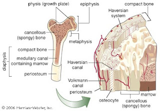
Bones are actually living organisms in the body that have nerves blood supply and cells. There is connective tissue inside the bone that surrounds the blood vessels. Cells as they are in all parts of the body and the driving force of the bone creating bone tissue, breaking down old bone tissue.

a. Fetal bone formation
When bones form in the fetus they start with cartilage. Cartilage is thought of as a type of bone but is technically a type of very dense connective tissue. Once the cartilage has formed bony tissue fills around the blood vessels. The new bone tissue is formed inside of the cartilage at the growth plate that lies between the diaphylis and epiphysis and the two ends of the bone. Then the final bone starts to form, this happens in three stages.
b. Medullary cavity
The Medullary cavity is located in the diaphysis and is a hollow area shaped like a tube. This is where the bone marrow is and where the blood cells of the bone are created.

c. Bone tissue what's so special about bone tissue?
Bone tissue is what supports our body without it and our muscles we’d be a big lump on the floor. Bones also protect the organs from the outside world. And bones are kind enough to store most of our calcium and phosphate.
6. Calcium regulation in the bones
Bones store calcium and are essential to the health of the bone. Calcium is also needed for cell metabolism and muscle cells. When bone loses calcium they become weaker. It is the role of the thyroid and parathyroid to regulate calcium in the body. When bones don’t get enough calcium osteoporosis is manifested. Osteoporosis is a disease that effects more women than men and levels are high in the elderly. The weakness of the bones can lead to soreness and easy fractures and breaking.
Sources:
http://www.ncbi.nlm.nih.gov/books/bv.fcgi?rid=cooper.section.1790, http://www.cytochemistry.net/Cell-biology/actin_filaments_intro.htm, http://www.mpimf-heidelberg.mpg.de/~holmes/muscle/muscle1.html, http://www.wisegeek.com/what-is-cartilage.htm, http://www.technion.ac.il/~mdcourse/274203/lect5.html, Frolich PowerPoint for cells and Human Biology 10th edition, Human Biology 10th
No comments:
Post a Comment